
3241 FUT Hair Transplant Case Study
[vc_row][vc_column width=”1/2″][vc_column_text]History: This 50 year old male had the jarring experience of looking in the mirror and seeing his Dad staring back at him. He was otherwise healthy and had enjoyed a lifetime of thick hair over the rest of his head, so he always thought he would be able to avoid hair loss. Additionally, he was comfortable with the fact that a normal mature male hairline has some temple recession, so the slow thinning that had been slowly progressive had not bothered him – up to this point. He had not ever tried any therapies for maintaining his hair. He hoped to simply fill in the corners and reframe his face with hair surgery.
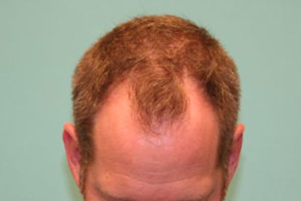
Physical Exam: This patient’s hair was a medium brown throughout and showed significant hair thinning at the fronto-temporal corners and in the vertex (crown) areas. The hairs were “miniaturized” on microscopic exam, except in the safe “donor” area at the back of his scalp, where the density was well above average. The rest of his physical exam was unremarkable. He kept his hair a medium length and anticipated that he would never choose to wear his hair short.
[/vc_column_text][/vc_column][vc_column width=”1/2″][vc_column_text]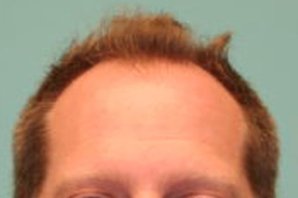
 [/vc_column_text][/vc_column][/vc_row][vc_row height=”auto”][vc_column][vc_column_text]Plan: After discussions about the importance of initial stabilizing his hair loss with medical therapy as a first priority, he chose to start finasteride 1mg per day and try minoxidil 5% foam once a day for at least a year to maintain his remaining hair. Since the fronto-temporal corners did not have any salvageable remaining hair, and since reframing his face was his primary goal, his long-term plan included filling in the frontal areas with hair grafts. He decided to obtain as many grafts as possible using the linear method of “strip” harvest since the multi-dotted diffuse scarring of the FUE method would provide him no cosmetic advantage.[/vc_column_text][us_separator size=”custom” height=”32px”][/vc_column][/vc_row][vc_row height=”auto”][vc_column][vc_column_text]
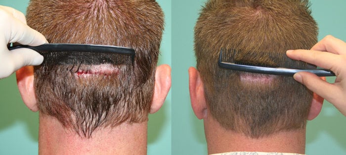
Donor area immediately after and 1 year post-op
Procedure: First, the “safe donor zone” was identified for this particular patient and the target hairline was drawn and agreed to. Then the area was numbed with a “ring block” that the patient described as “about 5 minutes of pinching and burning,” after which his head felt like “a helmet.” Since this gentleman chose to obtain grafts with a linear excision, a shallow (1mm) “score” was made behind both of his ears (parietal areas) through the area known as the “safe donor zone.” The hair was lifted out of place during the surgical part, and then allowed to fall back down and cover the area so that the donor area appeared completely undisturbed immediately after the procedure. He then laid on his back for the rest of the procedure and watched movies (or napped!).
This tissue was then observed under the microscope and the individual grafts (aka “follicular units”) were separated, counted, and categorized so that they could be transplanted in the ideal natural arrangement. For instance, single-haired follicular units (AKA “grafts”) were selected to rebuild the fine edge of the hairline, while two- and three-haired grafts were used to build density behind the hairline. Incisions were tiny and sized to fit the different graft sizes. Particular care was given to the direction and angulation of the sites. Grafts will tend to lift at a 15 degree “up” angle as they grow in, which means that in order to compensate and match existing hair, a slight “down” angle needs to be created. Hair grows out the way that the doctor makes the sites, so all of this technique is performed by the surgeon by hand in every patient and for every single graft. The full surgery lasted about 8 hours (~6.25 hours graft out of body time) with a break for lunch.[/vc_column_text][/vc_column][/vc_row][vc_row height=”auto”][vc_column][vc_column_text]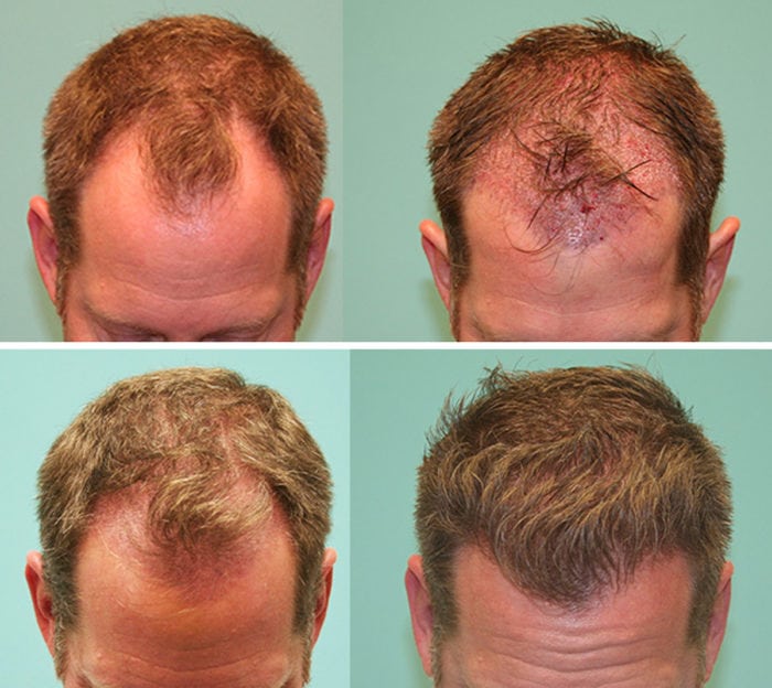
Results: This patient obtained 3241 grafts (407 singles 1593 doubles 1241 triples). He started finasteride a little over a month prior to surgery along with minoxidil, and experienced earlier than normal re-growth with significant improvement even at the three month mark (usually expected at about 6 months). Results shown here are at the 6 month post-operative visit and also at the 12 month visit. He continues to take both finasteride and use minoxidil daily.
Discussion: This case study demonstrates a hair transplant that is typical in many ways of the “state of the art.” A typical linear or “strip” harvest will have a high level of graft survival and a high yield, especially when handled by an experienced staff. In this case all the grafts were placed by hand with forceps (three staff members with 8-22 years of experience each!) into pre-made sites with the physician present at all times checking the graft quality and checking the handling of the hairs at every step of the way. FUE surgeries and surgical practices that use implanters can also give excellent results like this.
The key differentiating factor from a patient perspective is that practices that perform hair surgery daily can achieve and maintain this level of function more capably than an office which may perform a hair surgery less commonly. Excellent training, and years of experience will usually make a surgeon’s skill level comparatively higher, but although the surgeon is often touted as the “expert,” in reality it is the entire dedicated and experienced TEAM that makes a hair surgery of the highest quality. Further, individual patient characteristics, like the willingness to use medical means to improve hair, and factors like hair density, color, caliber, and curl etc. often make similar hair surgeries seem like one is more successful than another. In this case, the patient was extremely pleased with his results and did not require any additional surgery.[/vc_column_text][us_separator height=”50px” size=”custom”][/vc_column][/vc_row]